

TL;DR:
- Mast cell activation syndrome (MCAS) is diagnosed through three criteria: episodic multi-system symptoms, objective mediator elevation, and response to treatment. Proper timing and laboratory testing of mediators like tryptase are essential for confirmation. Recognizing triggers and working with specialists improve management and prognosis.
Mast cell activation syndrome (MCAS) is a disorder of inappropriate, episodic mast cell mediator release that causes multi-system allergic-type symptoms and requires three specific clinical criteria to diagnose: recurrent symptoms affecting at least two organ systems, objective laboratory evidence of elevated mast cell mediators using the 120% + 2 ng/mL tryptase rule, and a meaningful clinical response to medications that block or stabilize mast cells. Symptoms alone are not sufficient. The Cleveland Clinic and AAAAI both emphasize that objective mediator evidence is non-negotiable for a valid diagnosis. If you are experiencing anaphylaxis signs right now (throat swelling, severe drop in blood pressure, respiratory distress), use epinephrine and call 911 immediately. For recurrent multi-system episodes without an emergency, your next step is a referral to an allergist or immunologist who can coordinate timed mediator testing.
-
Three required criteria: multi-system episodic symptoms + objective mediator rise + response to anti-mast cell therapy
-
Key tryptase formula: event tryptase must exceed (1.2 × baseline) + 2 ng/mL
-
Emergency red flag: any sign of anaphylaxis requires intramuscular epinephrine and 911, not a wait-and-see approach
-
Specialist path: allergist/immunologist for suspected MCAS; hematology if clonal disease is suspected
Pro Tip: Ask your clinician to write standing orders for timed tryptase and urine mediator collection before your next episode. The diagnostic window for serum tryptase is roughly 30 minutes to 2 hours after symptom onset. A sample drawn four hours later is often uninformative.
What are mast cells, and how does MCAS differ from related conditions?
Mast cells are immune sentinels found in nearly every tissue, especially at barrier sites like the skin, gut lining, and airways. When they detect a threat, they release a cascade of chemical mediators: histamine, tryptase, prostaglandin D2 (PGD2), and leukotrienes. In a healthy immune response, that release is proportionate and targeted. In MCAS, mast cells fire inappropriately, releasing mediators in response to triggers that should not provoke a reaction, or releasing them without any identifiable trigger at all.
The Collegium Internationale Allergologicum 2022 update defines three distinct categories of MCAS, and the distinction genuinely changes what tests you need and what the long-term picture looks like:
-
Primary (clonal) MCAS: Mast cells carry a somatic mutation, most commonly KIT D816V. This form overlaps with systemic mastocytosis and requires bone marrow evaluation and genetic testing to characterize fully.
-
Secondary (reactive) MCAS: Mast cells are structurally normal but are chronically activated by an underlying driver, such as IgE-mediated allergy, autoimmune disease, or chronic infection.
-
Idiopathic MCAS: No clonal mutation and no identifiable secondary cause. This is the most common category in clinical practice, and it is also the most frequently overdiagnosed.
MCAS is not the same as systemic mastocytosis, which involves a clonal proliferation of mast cells in organs and carries its own diagnostic and prognostic criteria. It is also not a blanket explanation for chronic fatigue or unexplained pain. The 2022 Collegium update explicitly recommends investigating genetic contributors, including hereditary alpha-tryptasemia (a TPSAB1 gene copy number variant), when baseline tryptase is persistently elevated and symptoms are severe. Understanding which category applies to you shapes every subsequent decision, from which labs to order to whether a hematologist needs to be involved.
Why is MCAS so often missed or misdiagnosed?
MCAS is a diagnosis of exclusion. Symptoms are episodic, systemic, and nonspecific, which means they overlap with a long list of other conditions. Flushing, brain fog, and GI cramping can each be explained by half a dozen diagnoses. When they occur together and then resolve completely between episodes, the picture becomes more distinctive, but most clinicians still need to rule out other causes before MCAS can be confirmed.

The AAAAI Work Group Report on MCAS cautions explicitly against overdiagnosis, noting that objective mediator rises and therapeutic response are both required parts of confirmation. Symptoms alone have led to widespread misattribution in recent years, particularly on social media, where MCAS has been proposed as an explanation for nearly every chronic symptom cluster.
Conditions that must be ruled out before confirming MCAS:
-
IgE-mediated allergic disease (standard allergy testing)
-
Systemic mastocytosis (bone marrow biopsy, serum tryptase, KIT D816V)
-
Postural orthostatic tachycardia syndrome (POTS)
-
Hypermobile Ehlers-Danlos syndrome (hEDS)
-
Carcinoid tumor or VIPoma (urine 5-HIAA, vasoactive intestinal peptide)
-
Medication reactions (NSAIDs, opioids, contrast agents)
-
Functional GI disorders and histamine intolerance
-
Pheochromocytoma (urine catecholamines)
| Feature | MCAS | Systemic Mastocytosis | IgE-Mediated Allergy |
|---|---|---|---|
| Clonal mast cell mutation | Possible (primary type) | Yes (KIT D816V common) | No |
| Organ infiltration by mast cells | No | Yes | No |
| Elevated baseline tryptase | Sometimes | Usually | Rarely |
| Bone marrow involvement | No (unless clonal) | Yes | No |
| Responds to antihistamines | Often | Partially | Yes |
| Triggered by specific allergen | Sometimes | Sometimes | Yes (IgE-specific) |
The Merck Manual notes that MCAS frequently coexists with POTS and hypermobile EDS, which complicates the diagnostic picture further. Treating mast cell instability in these patients can meaningfully reduce their overall symptom burden, but it does not replace addressing the coexisting condition.
What MCAS symptoms look like across body systems
The hallmark of mast cell activation is that symptoms arrive in episodes, often within minutes of a trigger, and then resolve between flares. That episodic pattern is one of the most clinically useful clues. Chronic, unremitting symptoms that never fluctuate are less characteristic of MCAS.
Skin
-
Flushing (sudden redness, warmth, often without sweating)
-
Urticaria (hives) and angioedema
-
Dermatographism (skin writing: raised welts from light pressure)
-
Pruritus without visible rash
Gastrointestinal
-
Nausea, vomiting, abdominal cramping
-
Diarrhea or alternating bowel habits
-
Bloating and early satiety
Respiratory
-
Wheezing, chest tightness
-
Throat tightness or a sensation of swelling
-
Nasal congestion and rhinorrhea during episodes
Cardiovascular
-
Tachycardia and palpitations
-
Hypotension or near-syncope (especially after standing)
-
Flushing with a drop in blood pressure
Neurologic
-
Brain fog, difficulty concentrating
-
Headaches, often during or after episodes
-
Anxiety or a sense of impending doom (a recognized mediator effect, not purely psychological)
Ocular
-
Itchy, watery eyes
-
Conjunctival redness and swelling
-
Periorbital puffiness during flares
Red Flag: Hypotension, throat swelling, severe respiratory distress, or loss of consciousness during an episode are signs of anaphylaxis. Use epinephrine immediately and call 911. Do not wait to see if symptoms resolve on their own.
Isolated fatigue, chronic widespread pain, or cognitive difficulties that occur continuously and do not fluctuate with identifiable episodes are less likely to be caused by MCAS alone. Attributing these symptoms to mast cell disease without objective mediator evidence can delay the correct diagnosis.
What triggers mast cell episodes?
Mast cells in MCAS are, in a sense, hair-trigger alarms. Stimuli that would not provoke a reaction in a healthy person can set off a full mediator cascade. Triggers vary considerably between patients, which is why a personalized tracking approach matters far more than any generic avoidance list.
Common trigger categories:
-
Physical: Heat, cold, vibration, pressure, exercise, friction
-
Dietary: Alcohol, fermented foods, aged cheeses, shellfish, food additives (histamine-rich or histamine-releasing foods)
-
Medications: NSAIDs (aspirin, ibuprofen), opioids, radiocontrast media, certain antibiotics, neuromuscular blocking agents
-
Environmental: Strong fragrances, mold, pollens, latex, insect venoms
-
Emotional and physiologic: Acute stress, anxiety, hormonal fluctuations (many patients report worsening perimenstrually)
-
Infections: Viral or bacterial illness can destabilize mast cells and precipitate flares
Genetic factors can raise baseline susceptibility. Hereditary alpha-tryptasemia, caused by extra copies of the TPSAB1 gene, is associated with higher baseline tryptase, more severe anaphylaxis, and greater symptom burden. The KIT D816V mutation, relevant to primary MCAS and systemic mastocytosis, is a separate genetic consideration that a hematologist or allergist may test for when clonal disease is suspected.
Pro Tip: Keep a timestamped diary of every episode: what you ate, where you were, temperature, activity level, stress, and medications taken that day. After six to eight episodes, patterns usually emerge that neither you nor your clinician could have identified from memory alone.

How MCAS is diagnosed: tests, timing, and when to go further
The three diagnostic criteria are clear in principle but challenging in practice. You need recurrent multi-system symptoms, an objective mediator rise, and a documented response to anti-mast cell therapy. The hardest part is capturing the mediator rise, because elevations are transient and the diagnostic window is narrow.
The three required criteria:
-
Recurrent, episodic symptoms affecting two or more organ systems
-
Objective laboratory evidence of mast cell mediator elevation (tryptase using the 120% + 2 ng/mL formula, or validated urinary mediators)
-
Clinical response to medications that block or stabilize mast cells
| Test | What It Measures | Ideal Timing | Practical Notes |
|---|---|---|---|
| Serum tryptase (event) | Mast cell degranulation marker | Within 30 min–2 hours of symptom onset | Compare to baseline; apply 120% + 2 ng/mL rule |
| Serum tryptase (baseline) | Resting mast cell burden | Outside of any episode | Elevated baseline may suggest clonal disease |
| 24-hour urine N-methylhistamine | Histamine metabolite | Collected during/after symptomatic period | Refrigerate sample; high false-negative if collected outside event |
| 24-hour urine 11β-PGF2α | Prostaglandin D2 metabolite | During or after episode | Useful when tryptase is normal but symptoms are classic |
| Urinary leukotriene E4 (LTE4) | Leukotriene pathway activation | During or after episode | Supports diagnosis when other mediators are borderline |
| KIT D816V mutation | Clonal mast cell disease | Any time (blood or bone marrow) | Order when baseline tryptase is persistently elevated |
| TPSAB1 copy number | Hereditary alpha-tryptasemia | Any time (blood) | Consider when baseline tryptase >8 ng/mL without other cause |
False negatives are common when samples are delayed. A tryptase drawn three hours after symptom onset may be completely normal even in a true MCAS event. Coordinated sampling, ideally with written standing orders your clinician provides to a local ER or lab, is the single most effective way to improve diagnostic yield.
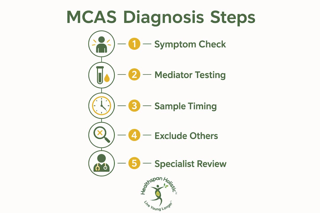
Bone marrow biopsy is not a routine part of MCAS workup. It becomes relevant when baseline tryptase is persistently elevated, when KIT D816V is detected in peripheral blood, or when clinical features suggest systemic mastocytosis. That decision belongs with a hematologist or experienced allergist.
When lab confirmation is elusive, the Merck Manual describes empirical therapeutic trials as a legitimate part of the diagnostic pathway. A supervised trial of H1 and H2 antihistamines, with or without a mast cell stabilizer, can provide meaningful diagnostic information when objective mediator evidence is borderline.
Timing callout: Serum tryptase is most informative within 30 minutes to 2 hours of onset. Urinary mediators require a 24-hour collection tied to a symptomatic period. Both samples collected outside these windows frequently produce false negatives.
How to treat mast cell activation: acute care and long-term management
Treatment for mast cell disorder management has two distinct phases: stopping an acute episode and preventing future ones. Most patients need both.
Acute management:
-
Epinephrine (intramuscular, autoinjector): First-line treatment for anaphylaxis. Every patient with a history of severe MCAS episodes should carry two autoinjectors and have a written anaphylaxis action plan.
-
Positioning: Lie flat with legs elevated if hypotension is present. Sitting upright can worsen a blood pressure drop.
-
Emergency care: After epinephrine, go to an ER. Biphasic reactions (a second wave of symptoms hours later) occur in a meaningful subset of anaphylaxis cases.
Chronic regimens (stepwise approach):
-
H1 antihistamines (cetirizine, loratadine, fexofenadine): First step for most patients. Non-sedating options are preferred for daytime use; some patients benefit from a sedating H1 agent at night.
-
H2 antihistamines (famotidine, ranitidine): Block histamine receptors in the GI tract; often added when GI symptoms are prominent or H1 alone is insufficient.
-
Mast cell stabilizers: Cromolyn sodium (oral formulation for GI symptoms; inhaled for respiratory) and ketotifen (available in some U.S. compounding pharmacies) reduce mediator release rather than just blocking its effects.
-
Leukotriene modifiers (montelukast): Useful when LTE4 is elevated or when respiratory symptoms dominate.
-
Aspirin: Can reduce PGD2-driven flushing in patients who tolerate it, but must be used cautiously. NSAIDs are a known trigger for some MCAS patients, so aspirin should only be introduced under clinician supervision with a test dose.
-
Omalizumab (Xolair): An anti-IgE biologic used for refractory cases where standard regimens provide insufficient control. Requires specialist oversight and is typically reserved for patients who have failed multiple medication classes.
The rationale for combination therapy is straightforward: antihistamines block the downstream effects of mediators already released, while stabilizers reduce how much gets released in the first place. Using both together addresses the problem from two directions. The AAAAI Work Group Report stresses that medication trials should be supervised and evaluated for objective symptom change, not simply continued indefinitely without reassessment.
Safety notes:
-
Corticosteroids are sometimes used for acute flares but are not appropriate for long-term management due to well-known systemic effects.
-
Pregnancy and breastfeeding require medication review with a specialist; some antihistamines and stabilizers have limited safety data in these populations.
-
Drug interactions are relevant: some antihistamines affect cytochrome P450 enzymes and can alter the metabolism of other medications.
Practical self-care steps that can reduce episode frequency
Medication manages the chemistry. Self-care manages the environment and lifestyle factors that feed into it. Both matter, and neither replaces the other.
Daily and episode-based checklist:
-
Keep a timestamped symptom and exposure diary (food, environment, activity, stress, medications)
-
Identify and avoid confirmed personal triggers, not just generic lists from online sources
-
Carry your epinephrine autoinjector at all times, including during exercise and travel
-
Wear a medical alert bracelet or carry a card listing your diagnosis, medications, and emergency instructions
-
Inform all healthcare providers (dentists, surgeons, anesthesiologists) of your diagnosis before procedures
-
Discuss any new medication, supplement, or herbal product with your clinician before starting it
Environmental and lifestyle adjustments:
-
Temperature regulation: avoid rapid shifts from hot to cold; use fans or cooling vests if heat is a trigger
-
Sleep: consistent sleep timing supports circadian regulation of immune function, including mast cell activity
-
Hydration: adequate fluid intake helps buffer cardiovascular symptoms like orthostatic hypotension
-
Exercise: graded, low-intensity activity is generally better tolerated than high-intensity bursts; work with your clinician to find a safe starting point
-
Stress reduction: mindfulness, breathing exercises, and structured rest periods can reduce physiologic stress triggers
Unsupervised supplement experiments are a real risk in this population. Some supplements contain histamine-releasing compounds or interact with medications. The immune support guide for adults from Healthspan Holistic offers a useful framework for thinking about evidence-aligned supplementation under professional guidance rather than trial-and-error.
Pro Tip: Write a one-page episode plan and give copies to your household members, your primary care provider, and your local ER. It should list your baseline medications, known triggers, your usual early symptoms, and the exact steps to take if you cannot communicate. This single document can prevent dangerous delays in emergency care.
Which specialists should you see, and how do you prepare?
Getting to the right specialist faster is one of the most practical things you can do to shorten the diagnostic timeline. The first referral depends on your dominant symptoms.
Who to see first:
-
Allergist/immunologist: The primary specialist for suspected MCAS. They can coordinate timed mediator testing, perform allergy evaluation to rule out IgE-mediated disease, and manage the medication regimen.
-
Hematologist: Consult when baseline tryptase is persistently elevated, KIT D816V is detected, or systemic mastocytosis is on the differential. Bone marrow biopsy, if needed, is typically performed and interpreted by hematology.
-
Cardiologist or autonomic specialist: Relevant when POTS or significant cardiovascular symptoms (syncope, sustained tachycardia) are present alongside suspected MCAS.
-
Gastroenterologist: Consider when GI symptoms are severe or when inflammatory bowel disease, eosinophilic GI disease, or carcinoid tumor needs to be excluded.
What to bring to your first appointment:
-
Symptom diary with dates, times, duration, and associated exposures
-
Photos or video of skin reactions (hives, flushing, angioedema) during episodes
-
Complete medication and supplement list, including OTC antihistamines you have already tried
-
Results from any prior allergy testing, blood work, or imaging
-
A written summary of your episode pattern: how often, how severe, how long they last, and what (if anything) makes them better
Tests the specialist may order:
-
Timed serum tryptase (event and baseline)
-
24-hour urine N-methylhistamine, 11β-PGF2α, and LTE4
-
KIT D816V mutation testing (peripheral blood, or bone marrow if indicated)
-
TPSAB1 copy number for hereditary alpha-tryptasemia
-
Standard allergy panel (skin prick testing or specific IgE)
-
Tilt table test or standing heart rate test if POTS is suspected
Ask the clinic directly how they handle rapid sampling during acute events. Some centers provide written instructions for local ERs or labs so that timed draws can happen even when you cannot get to the specialist’s office during a flare.
What is the long-term outlook for people with MCAS?
Most patients with idiopathic or secondary MCAS achieve meaningful symptom control with targeted therapy and trigger management. The condition does not typically shorten life expectancy in these categories, but it can substantially affect quality of life if left unmanaged or mismanaged.
Prognosis by category:
-
Idiopathic MCAS: Generally favorable with appropriate medication. Many patients stabilize over months to years, though some require ongoing multi-drug regimens.
-
Secondary MCAS: Prognosis depends heavily on the underlying driver. Treating the root cause (resolving an infection, managing autoimmune disease) often reduces mast cell instability.
-
Primary (clonal) MCAS: Requires monitoring for progression to systemic mastocytosis. Most patients with indolent systemic mastocytosis have a normal life expectancy, but periodic reassessment is warranted.
Potential complications to monitor:
-
Recurrent anaphylaxis, which carries cumulative risk with each episode
-
Osteoporosis, particularly in patients with systemic mastocytosis where mast cell mediators affect bone remodeling
-
Cardiovascular effects from chronic mediator exposure (tachycardia, blood pressure instability)
-
Psychological impact: anxiety, depression, and social withdrawal are common in patients with unpredictable, severe episodes
Follow-up schedule:
-
Review medication efficacy and tolerability every three to six months during the first year
-
Repeat mediator testing when symptoms change significantly or when a new trigger pattern emerges
-
Bone marrow re-evaluation in clonal cases is guided by the hematologist based on clinical trajectory
-
Multidisciplinary care (allergist, hematologist, cardiologist, mental health support) produces better outcomes than single-specialist management in complex cases
Getting accurate lab results: the testing workflow that actually works
The most common reason MCAS goes unconfirmed is not that the condition is absent. It is that the samples were drawn too late, handled incorrectly, or collected outside of a symptomatic period. Understanding the mechanics of the testing window is what separates a useful workup from a frustrating series of normal results.
| Test | Optimal Sample Timing | Relative Sensitivity | Practical Handling Notes |
|---|---|---|---|
| Serum tryptase (event) | 30 min–2 hours after onset | Highest within first hour | Serum separator tube; process promptly; freeze if not run same day |
| Serum tryptase (baseline) | Any time, outside episode | Reference standard | Must be drawn at least 24 hours after last episode |
| 24-hour urine N-methylhistamine | During/after symptomatic period | Moderate; improves with event capture | Refrigerate entire collection; avoid antihistamines prior if possible |
| 24-hour urine 11β-PGF2α | During/after symptomatic period | Moderate | Same collection protocol as N-methylhistamine |
| Urinary LTE4 | During/after symptomatic period | Variable | First morning void or 24-hour collection; avoid leukotriene modifiers before testing |
False negatives are the rule, not the exception, when samples are collected outside these windows. The AAAAI guidance on MCAS specifically highlights that coordinated sampling during flares is the critical variable for diagnostic sensitivity. A single normal tryptase drawn in a clinic two days after an episode tells you almost nothing.
Improving diagnostic yield comes down to three practical steps: pair every event tryptase with a baseline drawn outside of episodes, collect urine mediators during symptomatic periods rather than on arbitrary calendar days, and work with a specialty center or coordinated lab service that understands the timing requirements.
Pro Tip: Ask your allergist or immunologist for a written sampling protocol you can hand to an ER nurse or local lab technician. Include the exact tubes needed, the timing window, storage instructions, and the lab address where samples should be sent. Clinicians who have done this report a dramatic improvement in the quality of results they receive.
Integrated diagnostic programs that coordinate timed mediator sampling with advanced blood panels and longitudinal symptom tracking increase diagnostic yield compared with piecemeal testing. Healthspan Holistic’s inflammation blood test panels and AI-enabled analytics platform can help integrate mediator results with broader health data for a more complete picture, particularly in complex cases where multiple systems are involved.
Key Takeaways
Mast cell activation syndrome requires three objective criteria for diagnosis, not just symptoms, and timed mediator testing is the step most patients miss.
| Point | Details |
|---|---|
| Three criteria required | Diagnosis needs multi-system episodes, a mediator rise confirmed by validated laboratory criteria, and response to anti-mast cell therapy. |
| Timed sampling is critical | Serum tryptase must be drawn within 30 min–2 hours of onset; delayed samples frequently produce false negatives. |
| Anaphylaxis is a medical emergency | Throat swelling, hypotension, or severe respiratory distress require epinephrine and 911, not watchful waiting. |
| Specialist coordination speeds diagnosis | An allergist/immunologist is the first referral; hematology joins when clonal disease is suspected. |
| Healthspan Holistic supports the workup | Coordinated lab testing, AI analytics, and personalized coaching through Healthspan Holistic can integrate mediator panels with broader health data for a structured diagnostic and management plan. |
My take on MCAS
There is a particular kind of exhaustion that comes with a condition that looks invisible between episodes. You feel fine at the doctor’s office. Your labs come back normal because the sample was drawn on the wrong day. You get told it might be anxiety, or IBS, or stress. And then you have another episode that leaves you on the floor.
What I find most striking about MCAS is how much the diagnostic outcome depends on logistics rather than biology. The condition is real. The mediators are measurable. But capturing them requires a level of coordination that most healthcare systems are not set up to provide spontaneously. The patients who get diagnosed faster are almost always the ones who came prepared: a symptom diary, photos of their rashes, a list of every medication they had tried, and a clinician who understood why the timing of the blood draw mattered.
The frustration is valid. So is the persistence. Working methodically with a specialist, tracking episodes with precision, and pushing for timed mediator testing rather than accepting a string of inconclusive results is not being difficult. It is being your own best advocate in a diagnostic process that genuinely rewards preparation. The evidence-based path exists. It just takes patience and the right team to walk it.
- Chris
Healthspan Holistic can help you build a smarter diagnostic plan
For people navigating a complex, multi-system condition like MCAS, the difference between a frustrating diagnostic odyssey and a clear path forward often comes down to how well your testing is coordinated and interpreted.

The 90 Day Journey to Longevity is a structured program that combines testing, coaching, and a personalized health plan, making it a practical fit for anyone who wants to move beyond piecemeal lab work and build a longitudinal picture of their health. Whether you are in the early stages of a suspected MCAS workup or managing a confirmed diagnosis alongside other conditions, having a coordinated team and integrated data makes a measurable difference.
1st Time Customers can take advantage of our BUY 1 GET 1 50% OFF special offer on all supplements.
Useful sources
These are the primary references used throughout this article. Each one is vetted, peer-reviewed or society-endorsed, and appropriate for both patients and clinicians seeking deeper reading.
-
Mast Cell Activation Syndromes: Collegium Internationale Allergologicum Update 2022 — The most current peer-reviewed consensus on MCAS classification, diagnostic criteria, genetic contributors (KIT D816V, hereditary alpha-tryptasemia), and management. Essential reading for understanding the three MCAS categories.
-
AAAAI Mast Cell Disorders Committee Work Group Report (JACI, 2019) — The American Academy of Allergy, Asthma & Immunology’s formal diagnostic and management guidance. Covers validated mediators, stepwise diagnosis, and the caution against overdiagnosis.
-
AAAAI Patient Guidance on MCAS — Patient-facing resource from the AAAAI covering symptoms, testing logistics, and when to seek specialist care.
-
Cleveland Clinic: Mast Cell Activation Syndrome — Accessible clinical summary covering the tryptase formula, diagnostic requirements, and treatment overview. Useful for patients preparing for specialist appointments.
-
Merck Manual (Professional): Mastocytosis and Mast Cell Activation Syndrome — Clinician-level reference covering differential diagnosis, treatment classes, comorbidities (POTS, EDS), and empirical therapeutic trials.
-
University of Virginia GI Nutrition: Mast Cell Activation Syndrome Overview — Practical clinical overview with emphasis on GI manifestations, dietary triggers, and patient management strategies.
FAQ
How do you treat MCAS?
Treatment combines acute and chronic strategies: intramuscular epinephrine for anaphylaxis, and a stepwise chronic regimen of H1 and H2 antihistamines, mast cell stabilizers (cromolyn, ketotifen), leukotriene modifiers (montelukast), and omalizumab for refractory cases, all supervised by a specialist.
How do you know if you have mast cell activation syndrome?
Diagnosis requires three criteria: recurrent multi-system episodes, an objective mediator rise (serum tryptase meeting the 120% + 2 ng/mL formula, or elevated urinary mediators), and a clinical response to anti-mast cell therapy. Symptoms alone, without lab confirmation, are not sufficient for a valid diagnosis.
What are MCAS symptoms in the eye?
Ocular symptoms include itchy, watery eyes, conjunctival redness and swelling, and periorbital puffiness during episodes. These typically occur alongside symptoms in other systems rather than in isolation.
What conditions are similar to MCAS?
The closest overlapping conditions are systemic mastocytosis, IgE-mediated allergic disease, POTS, hypermobile Ehlers-Danlos syndrome, carcinoid tumor, pheochromocytoma, and histamine intolerance. All must be evaluated and excluded before MCAS is confirmed, since each requires a different treatment approach.
Leave a comment: